KMTC HEALTH DATA CLASSIFICATIONS NOTES
| Institution | Kenya Medical Training College |
| Course | HEALTH RECORDS |
| Year | 1st Year |
| Semester | Unknown |
| Posted By | Brian Mike |
| File Type | |
| Pages | 98 Pages |
| File Size | 1.57 MB |
| Views | 3695 |
| Downloads | 0 |
| Price: |
Buy Now
|
Description
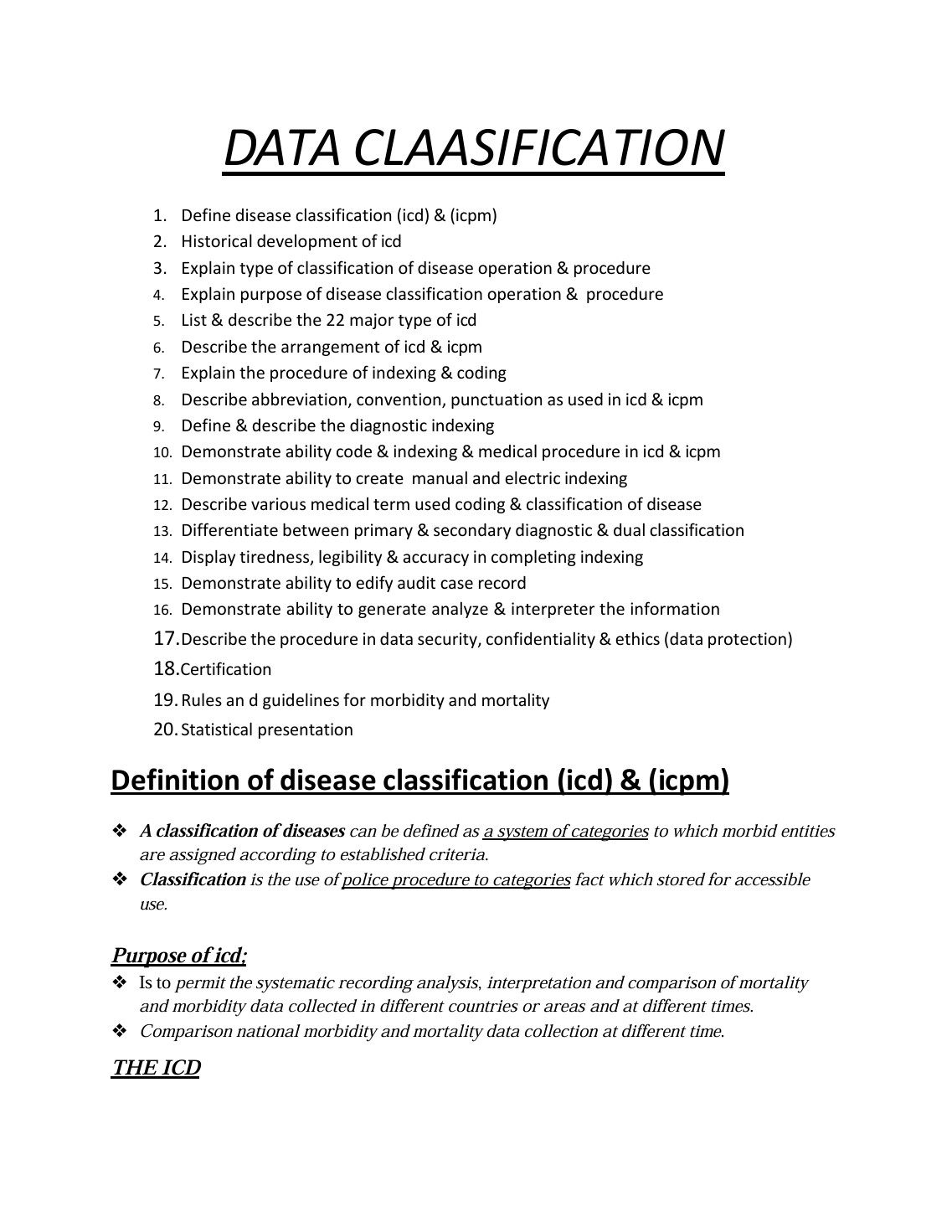
A classification of diseases can be defined as a system of categories to which morbid entities are assigned according to established criteria. Classification is the use of police procedure to categories fact which stored for accessible use.
Below is the document preview.
Introduction to ECG(Electrophysiology)
Trending!
The electrocardiogram is the graphical representation of the electrical activity of the heart
• It records the electrical activity of atrial ventricular cells in form of specific waveforms and complexes.
• The first ECG was introduced by Willem Einthoven, a Dutch physiologist, in the early 1900s
• ECG is an important diagnostic method in cardiac conditions.
27 Pages
2902 Views
0 Downloads
1.51 MB
Introduction to ECG (Basic Electrocardiogram)
Trending!
Indications For ECG Monitoring
Indicated for:-
• To monitor a patient’s heart rate and rhythm
• Evaluate the effects of disease or injury on heart function
• Evaluate pacemaker function
• Evaluate the response to medications
• Obtain a baseline recording before, during, and after a medical procedure
80 Pages
2490 Views
0 Downloads
3.72 MB
Junctional Rhythms ( Electrophysiology)
Trending!
The (AVN) Atrial Ventricular Node is a group of specialized cells located in the lower part of the right atrium above the tricuspid valve base. Its function is to Delay, Relay and Filter impulses as they pass through the Ventricles.
The nodal cells have pacemaker properties and can pace the heart at a rate of 40-60b/min. Rhythms coming from the AV junction are called .Junctional dysrhythmias. The electrical impulse must travel backward
( Retrograde) to activate the atria.
17 Pages
2470 Views
0 Downloads
624.95 KB
Sinus Mechanisms
Trending!
A normal heartbeat results from an electrical impulse that originates from the hearts primary pace maker-(SA node)
• The Normal sinus rhythm records the hearts electrical impulse that starts in the SAN spreading through the normal conduction pathway.
• The SAN dominates other areas that may pace the heart slower and abnormally.
All other rhythms will be compared to the Normal Sinus
Rhythm
44 Pages
2655 Views
0 Downloads
1.72 MB
Ventricular Rhythms
Trending!
The ventricles (purkinje fibres) may assume the function of pacing the heart, they pace at a slower rate 20-40b/min which cannot sustain the body's perfusion requirements. Rhythms originating from the ventricles are called ventricular arrhythmias because they originate in the ventricles.
Ventricular arrhythmias occur when:-
a) The SAN fails to initiate an impulse
b) The AVN does not pick to pace
c) There is an irritable foci in the ventricular muscle
61 Pages
2506 Views
0 Downloads
2.42 MB
Sickle cell Crisis
Trending!
Sickle cell disease (SCD)
• A group of hereditary disorders in which the normal adult hemoglobin (hemoglobin A) is partly or completely replaced by abnormal sickle hemoglobin (HgbS).
• The most common genetic hematologic condition in children
• Transmitted by autosomal recessive pattern of inheritance.
• Patient with this condition is homozygous for the sickle cell gene, i.e. both genes are abnormal.
• The basic defect responsible for the sickling of
erythrocytes is contained in the globin fraction of hemoglobin
• The mode of transmission is hereditary
• The gene that determines the production of HgbS is situated on an autosome.
47 Pages
2342 Views
0 Downloads
833.89 KB
Treatment modalities for hematological disorders
Trending!
These are therapies aimed at preventing, alleviating the underlying cause or treating the hematological disorder
• They include
1. Nutritional therapy: meal management to ensure rich sources of iron, vitamin K, vitamin B9.
2. Supplements: administration of folate, iron sulfate.
3. Treatment of underlying cause: antimalarial, deworming, ulcer treatment
21 Pages
2302 Views
0 Downloads
318.84 KB
Valvular heart disease
Trending!
Outline
1. Review the role of valves in cardiac cycle
2. Define valvular heart disease
3. Identify types of VHD
4. Explain the assessment and diagnostic tests
5. Describe the management of patient with VHD
46 Pages
2297 Views
0 Downloads
680.83 KB
Assessment and diagnostic evaluation of patient with haematological disorder
Trending!
History taking
1. Nutrition: feeding habits; typical meal – to determine deficiencies
2. use of prescription and over-the-counter medications: most hematological conditions can result from herbs, or certain medications.
3. Prior chemotherapy or radiotherapy
16 Pages
2300 Views
1 Downloads
372.41 KB
Disseminated intravascular coagulation (DIC)
Trending!
It is a condition of combine platelet and coagulation factor disorder.
• It is characterized by widespread coagulation and bleeding in the vascular compartment.
• DIC occurs secondary to inappropriate systemic activation of normal clotting mechanisms.
• It is associated with underlying disease manifested as uncontrolled activation of coagulation and fibrinolysis.
21 Pages
2646 Views
1 Downloads
309.13 KB